Cheney Mason would like the judge to explain to the jury why the faces have changes. JP> Do you want me to tell them the truth, (Chucl=kle) Cheny Mason> Something like that (chuckle) JP> Okay , I will tell them something.
The Jury is seated. The judge tells them that Dr Rodriquez will return on Monday. He makes no mention of Baez possible withholding information.
He did a second autopsy on Caylee Anthony.
Spitz is going to counter Dr G's examination.Spitz said he is questioning the Orlando medical examiner's neglect to open the child's skull during the original autopsy. Spitz said he has seen cases solved due to opening the skull.
He is stating his background and eductatio
n.
Medical School:
Hadassah Medical School, The Hebrew University Of Jerusalem
Jerusalem, Israel
Graduated: 1953
Residency Hospitals:
Hebrew University Hadassah Med School
Year completed: 1959
Tel Hashomer Govt Hospital
Year completed: 1957
Internship Hospital:
Tel Hashomer Govt Hospital
Year completed: 1955
Fellowship Hospital:
Med Exam Ofc
Baltimore, MD, USA
Year completed: 1961
He lives in Michigan and is a forensic pathologist. Spitz testified about his education, memberships and awards.
He was the medical examiner for Wayne County, Michigan. He has been on the editorial board for a number of American and European journals. He grew up speaking German because he was born in Germany, but he writes and lectures in German, French, Swiss and Hebrew. He is a professor of pathology at Wayne State University Medical School. He's published 14 articles related to drowning because it's been an area of great interest to him and several other pathologists. Michigan is a shoreline state so they have many drownings. He's written a text book about forensic pathology that is circulated worldwide. It's now in its fourth edition and he's working on a fifth edition. He's served as a lecturer for several groups and organizations worldwide. He worked on the assassination of President Kennedy and testified in the civil case against O.J. Simpson. He also testified in the Jon Benet Ramsey case in Boulder, Colorado and in the Mary Jo Kopechne civil case against Ted Kennedy. He has testified as an expert in all 50 states and Canada. Asked if he remembered how many times he's been qualified to testify as an expert in court he replied, "To tell the truth, I can't remember a time when I wasn't qualified". He was board certified in 1965 and has remained certified. He's done an estimated 60,000 autopsies in his career, although he does fewer now. He's licensed to practice medicine in many states, the District of Columbia and "all of the European countries." His hourly rate is $400 which is for travel time (he hires a driver), and he charges $5000 per day.
Spitz said determining the cause of death is very important. Did the victim drown in a lake or did he have a heart attack in the lake? The manner of death is classified in one of five categories; homicide, suicide, natural, accidental and undetermined. per FreeRyanWidmer.com
He has testifed in the many high profile cases such as, Mary Jo Kopeckne, Jon Benet Ramsey, and Phil Spector .
Dr Spitz is accepted as expert witness. He testifies Baez notified him. He was notified when the remains was found. There had not been an autopsy yet. He begins to testify he asked to be there but a sidebar is called by Ashton.
 |
| Cheney Mason, Defense Attorney. |
Cheney Mason questions witness.
He testifies he was not allowed to attend autopsy. He testifies it would have been helpful to see the state of the remains. Two forensic pathologist would have been helpful to review as autopsy is performed. It is common for forensic pathologist to consult and see if the concur on findings. "The request was denied."
When the official autopsy was completed he was given access to remains. He went to the Bryant Funeral home prior to cremation and conducted autopsy . DR G had not completed report.
 |
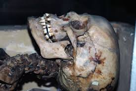
| View inside of a skull after top of skull removed. |
 |
| The top of a skull removed for autopsy |
He came with all kind of equipment , he thought the autopsy would have done but it was not done. The skull had not been opened. It should have been done. He opened the skull. Mason hands him a black cloth bag iwith a skull. It is a skull of an adult human being. It is real.He demonstrates how he "opened" the skull He sawed the roof of the skull from the rest. It is much like you would cut a pumpkin. Casey is tearful and turns away. Dr Werner steps down from stand for demonstration.
Cheney> What is remarkable about the contents you have WS.> He is explaining with a pic . He is reviewing the skull of Caylee Anthony.
Spitz is explaining black dots on skull. Jury continues to watch him. He is stating how you can tell how the skull was laying. He is outlining the decomposition pattern of Caylee's brain. The left side of brain decomposed last. He can tell by the dark spots inside the skull.
He says her brain essentially disappears but the remnants show she was laid on her left side ( put your left ear down)
 |
| Skull on Left side. Dr Werner Spitz testifies skull shows Caylee was laying on Left side when she decomposed. |
 |
| Caylee's skull was reported to be found in this position |
He states Caylees body was on it's left side, looking upward. This is important because the pics of Caylees skull at the remains site, was looking upward with the back of skull on the ground. This evidence infers Caylee
Mason>What deficiencies did you find in her (Dr.G) report. WS> The skull was not opened so I opened it. There was no discoloration of the skull. Like near the ear or back of the lower jaw. There were no fractures. The skull was intact. There was damage to long bones from animals. One long bone was opened by prior autopsy. I thought they had opened it to get access to bone marrow for testing. Mason> Did you read Dr G's report on the duct tape WS> The duct tape was loosely on the skull. There was no soft tissue. He prepared with gloves , gowns and aprons expecting to find soft tissue. There was nothing. He could touch with his hands. The bones were clean. The duct tape was not present. The duct tape was on the Left side and hanging on hair and vegetative roots. He states there is nothing to prove that the duct tape was on face nor is there anything on the bone to suggest it had duct tape. It appears the duct tape was there to keep the jaw in place. He demonstrates how without the duct tape the jaw would have fell off. Casey is crying.
WS states he would have expected DNA to be present on duct tape.
Spitz says Caylee's cause of death is unknown. WS states he put duct tape on his arm and removed it and there was hair with roots on tape. He states decomposition always is slower where there is pressure, like against a wall..In his opinion , duct tape placement was a later event not an early event.
Adipocere. WS is explaining it. He describes it as "soap like." There was no adipocere.
Adipocere ( /ˈædɨpɵsɪər/), also known as corpse, grave or mortuary wax, is a wax-like organic substance formed by the anaerobic bacterial hydrolysis of fat in tissue, such as body fat in corpses. In its formation, putrefaction is replaced by a permanent firm cast of fatty tissues, internal organs and the face.Adipocere is a crumbly, waxy, water-insoluble material consisting mostly of saturated fatty acids. Depending on whether it was formed from white or brown body fat, adipocere is grayish white or tan in color.[1] per Wilkipedia.
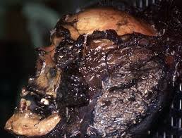 |
| Example Of Adipocere On Skull |
He reviewed Dr. G reports. He states cause of death unknown. You can not rule out accidental death.
Manner of death, homicide, suicide, natural death, accidental death. There are only four manners.
WS has perform and/ or assisted with 60, 000 autopsies.He has been a forensic pathologist for 60 years.
Recess.
Prosecutor Ashton crosses.
WS spoke to Baez , Mason and the Anthony family. WS read police reports but can not testify to which ones.
Spitz says he also went to the scene where Caylees' remains were found
WS understands that there was a month between the disappearance of child and the time reported. Everything needs to be evaluated together and not independently. Everything needs to fit together like a jigsaw puzzle. Aston> what do you know the background of case since WS testifies one needs to know all the facts in case to make a determination of means of death.
WS testifies that he understands the events when she last was seen alive was that she was taken to a babysitter. He does not know that he knows about facts of the case. Ashton> Dr G had considerably more information of case. WS> Whatever was in the report I read. (He was not told about pool drowning)
Ashton> One needs to know the predicates of the case. WS> We read the police reports. I did not say I did not read the reports. I remember the pertinent facts of the case. I do not recall what happened to her prior to the disappearance. I knew she was a healthy 3 year old child.
Ashton> What do you know or find significant?
WS> There was a pool at the house which could lead to a possibility of drowning. A month elapsed from disappearance to report. That is all I remember.
Ashton> DR. G not opening the skull was a violation of protocol
WS> In a high level case, we open the skull. The skull is part of the body. The head is extremely important. There is a protocol but I don't remember where. I was part of the committee that developed the protocol. To not open the head is a failure. Every forensic pathologist would tell you that to not open the head is "shoddy."
Ashton> In traditional autopsies , the skull is open to examine the skull
WS> NO it is one of the cases. He goes on to explain on why the skull is open. He would have not been able to tell you the position in which the body was laying if he did not open the skull.
Ashton> Certain protocols are for non skeletonized remains and skeletanized remains.
WS> NO.
Ashton> Shows WS a book in which he co-authored along with Dr Haskall and others. Show me where in the book there is a protocal for opening the skull.
WS> This is not a book on how to do an autopsy but how to determine results.
Ashton> He is pushing for a protocal. Mason> Objection, bolstering, JP> Overruled.
WS>THIS IS NOT A BOOK ON PROTCALS. IT IS NOT GOING TO SAY TO OPEN THE SKULL.
It is going to tell you the findings in the head in which the head would need to be open.
Ashton,> Are you familiar with the Minnesota protocal.....for disinterment and examination of remains.
http://www1.umn.edu/humanrts/instree/executioninvestigation-91.html (LINK)
A. Introduction
B. Proposed model skeletal analysis protocol
1. Scene investigation
2. Laboratory analysis of skeletal remains
3. Final report
4. Repository for evidence
2. Autopsy
The following Protocol should be followed during the autopsy:
(a) Record the date, starting and finishing times, and place of the autopsy (a complex autopsy may take as long as an entire working day);
(b) Record the name(s) of the prosecutor(s), the participating assistant(s), and all other persons present during the autopsy, including the medical and/or scientific degrees and professional, political or administrative affiliations(s) of each. Each person's role in the autopsy should be indicated, and one person should be designated as the principal prosector who will have the authority to direct the performance of the autopsy. Observers and other team members are subject to direction by, and should not interfere with, the principal prosector. The time(s) during the autopsy when each person is present should be included. The use of a "sign-in" sheet is recommended;
(c) Adequate photographs are crucial for thorough documentation of autopsy findings:
(i) Photographs should be in colour (transparency or negative/ print), in focus, adequately illuminated, and taken by a professional or good quality camera. Each photograph should contain a ruled reference scale, an identifying case name or number, and a sample of standard grey. A description of the camera (including the lens "f-number" and focal length), film and the lighting system must be included in the autopsy report. If more than one camera is utilized, the identifying information should be recorded for each. Photographs should also include information indicating which camera took each picture, if more than one camera is used. The identity of the person taking the photographs should be recorded;
(ii) Serial photographs reflecting the course of the external examination must be included. Photograph the body prior to and following undressing, washing or cleaning and shaving;
(iii) Supplement close-up photographs with distant and/or immediate range photographs to permit orientation and identification of the close-up photographs;
(iv) Photographs should be comprehensive in scope and must confirm the presence of all demonstrable signs of injury or disease commented upon in the autopsy report;
(v) Identifying facial features should be portrayed (after washing or cleaning the body), with photographs of a full frontal aspect of the face, and right and left profiles of the face with hair in normal position and with hair retracted, if necessary, to reveal the ears;
(d) Radiograph the body before it is removed from its pouch or wrappings. X-rays should be repeated both before and after undressing the body. Fluoroscopy may also be performed. Photograph all X-ray films;
(i) Obtain dental X-rays, even if identification has been established in other ways;
(ii) Document any skeletal system injury by X-ray. Skeletal X-rays may also record anatomic defects or surgical procedures. Check especially for fractures of the fingers, toes and other bones in the hands and feet. Skeletal X-rays may also aid in the identification of the deceased, by detecting identifying characteristics, estimating age and height, and determining sex and race. Frontal sinus films should also be taken, as these can be particularly useful for identification purposes;
(iii) Take X-rays in gunshot cases to aid in locating-the projectile(s). Recover, photograph and save any projectile or major projectile fragment that is seen on an X-ray. Other radio-opaque objects (pacemakers, artificial joints or valves, knife fragments etc.) documented with X-rays should also be removed, photographed and saved;
(iv) Skeletal X-rays are essential in children to assist in determining age and developmental status;
(e) Before the clothing is removed, examine the body and the clothing. Photograph the clothed body. Record any jewellery present;
(f) The clothing should be carefully removed over a clean sheet or body pouch. Let the clothing dry if it is bloody or wet. Describe the clothing that is removed and label it in a permanent fashion. Either place the clothes in the custody of a responsible person or keep them, as they may be useful as evidence or for identification;
(g) The external examination, focusing on a search for external evidence of injury is, in most cases, the most important portion of the autopsy;
(i) Photograph all surfaces - 100 per cent of the body area. Take good quality, well-focused, colour photographs with adequate illumination;
(ii) Describe and document the means used to make the identification. Examine the body and record the deceased's apparent age, length, weight, sex, head hair style and length, nutritional status, muscular development and colour of skin, eyes and hair (head, facial and body);
(iii) In children, measure also the head circumference, crown-rump length and crown-heel length;
(iv) Record the degree, location and fixation of rigor and livor mortis;
(v) Note body warmth or coolness and state of preservation; note any decomposition changes, such as skin slippage. Evaluate the general condition of the body and note adipocere formation, maggots, eggs or anything else that suggests the time or place of death;
(vi) With all injuries, record the size, shape, pattern, location (related to obvious anatomic landmarks), colour, course, direction, depth and structure involved. Attempt to distinguish injuries resulting from therapeutic measures from those unrelated to medical treatment. In the description of projectile wounds, note the presence or absence of soot, gunpowder, or singeing. If gunshot residue is present, document it photographically and save it for analysis. Attempt to determine whether the gunshot wound is an entry or exit wound. If an entry wound is present and no exit wound is seen, the projectile must be found and saved or accounted for. Excise wound tract tissue samples for microscopic examination. Tape together the edges of knife wounds to assess the blade size and characteristics;
(vii) Photograph all injuries, taking two colour pictures of each, labelled with the autopsy identification number on a scale that is oriented parallel or perpendicular to the injury. Shave hair where necessary to clarify an injury, and take photographs before and after shaving. Save all hair removed from the site of the injury. Take photographs before and after washing the site of any injury. Wash the body only after any blood or material that may have come from an assailant has been collected and saved;
(viii) Examine the skin. Note and photograph any scars, areas of keloid formation, tattoos, prominent moles, areas of increased or decreased pigmentation, and anything distinctive or unique such as birthmarks. Note any bruises and incise them for delineation of their extent. Excise them for microscopic examination. The head and genital area should be checked with special care. Note any injection sites or puncture wounds and excise them to use for toxicological evaluation. Note any abrasions and excise them; microscopic sections may be useful for attempting to date the time of injury. Note any bite marks; these should be photographed to record the dental pattern, swabbed for saliva testing (before the body is washed) and excised for microscopic examination. Bite marks should also be analysed by a forensic odontologist, if possible. Note any burn marks and attempt to determine the cause (burning rubber, a cigarette, electricity, a blowtorch, acid, hot oil etc.). Excise any suspicious areas for microscopic examination, as it may be possible to distinguish microscopically between burns caused by electricity and those caused by heat;
(ix) Identify and label any foreign object that is recovered, including its relation to specific injuries. Do not scratch the sides or tip of any projectiles. Photograph each projectile and large projectile fragment with an identifying label, and then place each in a sealed, padded and labelled container in order to maintain the chain of custody;
(x) Collect a blood specimen of at least 50 cc from a subclavian or femoral vessel;
(xi) Examine the head and external scalp, bearing in mind that injuries way be hidden by the hair. Shave hair where necessary. Check for fleas and lice, as these way indicate unsanitary conditions prior to death. Note any alopecia as this may be caused by malnutrition, heavy metals (e.g. thallium), drugs or traction. Pull, do not cut, 20 representative head hairs and save them, as hair may also be useful for detecting some drugs and poisons;
(xii) Examine the teeth and note their condition. Record any that are absent, loose or damaged, and record all dental work (restorations, fillings etc.), using a dental identification system to identify each tooth. Check the gums for periodontal disease. Photograph dentures, if any, and save them if the decedent's identity is unknown. Remove the mandible and maxilla if necessary for identification. Check the inside of the mouth and note any evidence of trauma, injection sites, needle marks or biting of the lips, cheeks or tongue. Note any articles or substances in the mouth. In cases of suspected sexual assault, save oral fluid or get a swab for spermatozoa and acid phosphatase evaluation. (Swabs taken at the tooth-gum junction and samples from between the teeth provide the best specimens for identifying spermatozoa.) Also take swabs from the oral cavity for seminal fluid typing. Dry the swabs quickly with cool, blown air if possible, and preserve them in clean plain paper envelopes. If rigor mortis prevents an adequate examination, the masseter muscles may be cut to permit better exposure;
(xiii) Examine the face and note if it is cyanotic or if petechiae are present;
a. Examine the eyes and view the conjunctiva of both the globes and the eyelids. Note any petechiae in the upper on lower eyelids. Note any scleral icterus. Save contact lenses, if any are present. Collect at least 1 ml of vitreous humor from each eye;
b. Examine the nose and ears and note any evidence of trauma, haemorrhage or other abnormalities. Examine the tympanic membranes;
(xiv) Examine the neck externally on all aspects and note any contusions, abrasions or petechia. Describe and document injury patterns to differentiate manual, ligature and hanging strangulation. Examine the neck at the conclusion of the autopsy, when the blood has drained out of the area and the tissues are dry;
(xv) Examine all surfaces of the extremities: arms, forearms, wrists, hands, legs and feet, and note any "defence" wounds. Dissect and describe any injuries. Note any bruises about the wrists or ankles that may suggest restraints such as handcuffs or suspension. Examine the medial and lateral surfaces of the fingers, the anterior forearms and the backs of the knees for bruises;
(xvi) Note any broken or missing fingernails. Note any gunpowder residue on the hands, document photographically and save it for analysis. Take fingerprints in all cases. If the decedent's identity is unknown and fingerprints cannot be obtained, remove the "glove" of the skin, if present. Save the fingers if no other means of obtaining fingerprints is possible. Save finger nail clippings and any under-nail tissue (nail scrapings). Examine the fingernail and toenail beds for evidence of object having been pushed beneath the nails. Nails can be removed b, dissecting the lateral margins and proximal base, and then the undersurface of the nails can be inspected. If this is done, the hands must be photographed before and after the nails are removed. Carefully examine the soles of the feet, noting any evidence of beating. Incise the soles to delineate the extent of any injuries. Examine the palms and knees, looking especially for glass shards or lacerations;
(xvii) Examine the external genitalia and note the presence of any foreign material or semen. Note the size, location and number of any abrasions or contusions. Note any injury to the inner thighs or peri-anal area. Look for peri-anal burns;
(xviii) In cases of suspected sexual assault, examine all potentially involved orifices. A speculum should be used to examine the vaginal walls. Collect foreign hair by combing the pubic hair. Pull and save at least 20 of the deceased's own pubic hairs, including roots. Aspirate fluid from the vagina and/or rest, for acid phosphatase, blood group and spermatozoa evaluation. Take swabs from the same areas for seminal fluid typing. Dry the swabs quickly with cool, blown air if possible, and preserve them in clean plain paper envelopes;
(xix) The length of the back, the buttocks and extremities including wrists and ankles must be systematically incised to look for deep injuries. The shoulders, elbows, hips and knee joints must also be incised to look for ligamentous injury;
(h) The internal examination for internal evidence of injury should clarify and augment the external examination;
(i) Be systematic in the internal examination. Perform the examination either by body regions or by systems, including the cardiovascular, respiratory, biliary, gastrointestinal, reticuloendothelial, genitourinary, endocrine, musculoskeletal, and central nervous systems. Record the weight, size, shape, colour and consistency of each organ, and note any neoplasia, inflammation, anomalies, haemorrhage, ischemia, infarcts, surgical procedures or injuries. Take sections of normal and any abnormal areas of each organ for microscopic examination. Take samples of any fractured bones for radiographic and microscopic estimation of the age of the fracture;
(ii) Examine the chest. Note any abnormalities of the breasts. Record any rib fractures, noting whether cardiopulmonary resuscitation was attempted. Before opening, check for pneumothoraces. Record the thickness of subcutaneous fat. Immediately after opening the chest, evaluate the pleural cavities and the pericardial sac for the presence of blood or other fluid, and describe and quantify any fluid present. Save any fluid present until foreign objects are accounted for. Note the presence of air embolism, characterized by frothy blood within the right atrium and right ventricle. Trace any injuries before removing the organs. If blood is not available at other sites, collect a sample directly from the heart. Examine the heart, noting degree and location of coronary artery disease or other abnormalities. Examine the lungs, noting any abnormalities;
(iii) Examine the abdomen and record the amount of subcutaneous fat. Retain 50 grams of adipose tissue for toxicological evaluation. Note the interrelationships of the organs. Trace any injuries before removing the organs. Note any fluid or blood present in the peritoneal cavity, and save it until foreign objects are accounted for. Save all urine and bile for toxicologic examination;
(iv) Remove, examine and record the quantitative information on the liver, spleen, pancreas, kidneys and adrenal glands. Save at least 150 grams each of kidney and liver for toxicological evaluation. Remove the gastrointestinal tract and examine the contents. Note any food present and its degree of digestion. Save the contents of the stomach. If a more detailed toxicological evaluation is desired, the contents of other regions of the gastrointestinal tract may be saved. Examine the rectum and anus for burns, lacerations or other injuries. Locate and retain any foreign bodies present. Examine the aorta, inferior vena cava and iliac vessels;
(v) Examine the organs in the pelvis, including ovaries, fallopian tubes, uterus, vagina, testes, prostate gland, seminal vesicles, urethra and urinary bladder. Trace any injuries before removing the organs. Remove these organs carefully so as not to injure them artifactually. Note any evidence of previous or current pregnancy, miscarriage or delivery. Save any foreign objects within the cervix, uterus, vagina, urethra or rectum;
(vi) Palpate the head and examine the external and internal surfaces of the scalp, noting any trauma or haemorrhage. Note any skull fractures. Remove the calvarium carefully and note epidural and subdural haematomas. Quantify, date and save any haematomas that are present. Remove the dura to examine the internal surface of the skull for fractures. Remove the brain and note any abnormalities. Dissect and describe any injuries. Cerebral cortical atrophy, whether focal or generalized, should be specifically commented upon;
(vii) Evaluate the cerebral vessels. Save at least 150 grams of cerebral tissue for toxicological evaluation. Submerge the brain in fixative prior to examination, if this is indicated;
(viii) Examine the neck after the heart and brain have been removed and the neck vessels have been drained. Remove the neck organs, taking care not to fracture the hyoid bone. Dissect and describe any injuries. Check the mucosa of the larynx, pyriform sinuses and esophagus, and note any petechiae, edema or burns caused by corrosive substances. Note any articles or substances within the lumina of these structures. Examine the thyroid gland. Separate and examine the parathyroid glands, they are readily identifiable;
(ix) Dissect the neck muscles, noting any haemorrhage. Remove all organs, including the tongue. Dissect the muscles from the bones and note any fractures of the hyoid bone or thyroid or cricoid cartilages;
(x) Examine the cervical, thoracic and lumbar spine. Examine the vertebrae from their anterior aspects and note any fractures, dislocations, compressions or haemorrhages. Examine the vertebral bodies. Cerebrospinal fluid may be obtained if additional toxicological evaluation is indicated;
(xi) In cases in which spinal injury is suspected, dissect and describe the spinal cord. Examine the cervical spine anteriorly and note any haemorrhage in the paravertebral muscles. The posterior approach is best for evaluating high cervical injuries. Open the spinal canal and remove the spinal cord. Make transverse sections every 0.5 cm and note any abnormalities;
(i) After the autopsy has been completed, record which specimens have been saved. Label all specimens with the name of the deceased, the autopsy identification number, the date and time of collection, the name of the prosector and the contents. Carefully preserve all evidence and record the chain of custody with appropriate release forms;
(i) Perform appropriate toxicologic tests and retain portions of the tested samples to permit retesting;
a. Tissues: 150 grams of liver and kidney should be saved routinely. Brain, hair and adipose tissue may be saved for additional studies in cases where drugs, poisons or other toxic substances are suspected;
b. Fluids: 50 cc (if possible) of blood (spin and save serum in all or some of the tubes), all available urine, vitreous humor and stomach contents should be saved routinely. Bile, regional gastrointestinal tract contents and cerebrospinal fluid should be saved in cases where drugs, poisons or toxic substances are suspected. Oral, vaginal and rectal fluid should be saved in cases of suspected sexual assault;
(ii) Representative samples of all major organs, including areas of normal and any abnormal tissue, should be processed histologically and stained with hematoxylin and eosin (and other stains as indicated). The slides, wet tissue and paraffin blocks should be kept indefinitely;
(iii) Evidence that must be saved includes:
a. All foreign objects, including projectiles, projectile fragments, pellets, knives and fibres. Projectiles must be subjected to ballistic analysis;
b. All clothes and personal effects of the deceased, worn by or in the possession of the deceased at the time of death;
c. Fingernails and under nail scrapings;
d. Hair, foreign and pubic, in cases of suspected sexual assault;
e. Head hair, in cases where the place of death or location of the body prior to its discovery may be an issue;
(j) After the autopsy, all unretained organs should be replaced in the body, and the body should be well embalmed to facilitate a second autopsy in case one is desired at some future point;
(k) The written autopsy report should address those items that are emphasized in boldface type in the protocol. At the end of the autopsy report should be a summary of the findings and the cause of death. This should include the prosector's comments attributing any injuries to external trauma, therapeutic efforts, postmortem change, or other causes. A full report should be given to the appropriate authorities and to the deceased's family.
V. MODEL PROTOCOL FOR DISINTERMENT AND ANALYSIS OF SKELETAL REMAINS
A. Introduction
This proposed model protocol for the disinterment and analysis of skeletal remains includes a comprehensive checklist of the steps in a basic forensic examination. The objectives of an anthropological investigation are the same as those of a medicolegal investigation of a recently deceased person. The anthropologist must collect information that will establish the identity of the deceased, the time and place of death, the cause of death and the manner or mode of death (homicide, suicide, accident or natural). The approach of the anthropologist differs, however, because of the nature of the material to be examined. Typically, a prosector is required to examine a body, whereas an anthropologist is required to examine a skeleton. The prosector focuses on information obtained from soft tissues, whereas the anthropologist focuses on information from hard tissues. Since decomposition is a continuous process, the work of both specialists can overlap. An anthropologist may examine a fresh body when bone is exposed or when bone trauma is a factor. An experienced prosector may be required when mummified tissues are present. In some circumstances, use of both this protocol and the model autopsy protocol may be necessary to yield the maximum information. The degree of decomposition of the body will dictate the type of investigation and, therefore, the protocol(s) to be followed.
The questions addressed by the anthropologist differ from those pursued in a typical autopsy. The anthropological investigation invests more time ant attention to basic questions such as the following:
(a) Are the remains human?
(b) Do they represent a single individual or several?
(c) What was the decedent's sex, race, stature, body weight, handedness and physique?
(d) Are there any skeletal traits or anomalies that could serve to positively identify the decedent?
The time, cause and manner of death are also addressed by the anthropologist, but the margin of error is usually greater than that which can be achieved by an autopsy shortly after death.
This model protocol may be of use in many diverse situations. Its application may be affected, however, by poor conditions, inadequate financial resources or lack of time. Variation from the protocol may be inevitable or even preferable in some cases. It is suggested, however, that any major deviations, with the supporting reasons, should be noted in the final report.
B. Proposed model skeletal analysis protocol
1. Scene investigation
A burial recovery should be handled with the same exacting care given to a crime-scene search. Efforts should be co-ordinated between the principal investigator and the consulting physical anthropologist or archaeologist. Human remains are frequently exhumed by law enforcement officers or cemetery workers unskilled in the techniques of forensic anthropology. Valuable information may be lost in this manner and false information is sometimes [----- Page 35 of original----] generated. Disinterment by untrained persons should be prohibited. The consulting anthropologist should be present to conduct or supervise the disinterment. Specific problems and procedures accompany the excavation of each type of burial. The amount of information obtained from the excavation depends on knowledge of the burial situation and judgement based on experience. The final report should include a rationale for the excavation procedure.
The following procedure should be followed during disinterment:
(a) Record the date, location, starting and finishing times of the disinterment, and the names of all workers;
(b) Record the information in narrative form, supplemented by sketches and photographs;
(c) Photograph the work area from the same perspective before work begins and after it ends every day to document any disturbance not related to the official procedure;
(d) In some cases, it is necessary to first locate the grave within a given area. There are numerous methods of locating graves, depending on the age of the grave:
(i) An experienced archaeologist may recognize clues such as changes in surface contour and variation in local vegetation;
(ii) A metal probe can be used to locate the less compact soil characteristics of grave fill;
(iii) The area to be explored can be cleared and the top soil scraped away with a flat shovel. Graves appear darker than the surrounding ground because the darker topsoil has mixed with the lighter subsoil in the grave fill. Sometimes a light spraying of the surface with water may enhance a grave's outline;
(e) Classify the burial as follows:
(i) Individual or commingled. A grave may contain the remains of one person buried alone, or it may contain the commingled remains of two or more persons buried either at the same time or over a period of time;
(ii) Isolated or adjacent. An isolated grave is separate from other graves and can be excavated without concern about encroaching upon another grave. Adjacent
graves, such as in a crowded cemetery, require a different excavation technique because the wall of one grave is also the wall of another grave;
(iii) Primary or secondary. A primary grave is the grave in which the deceased is first placed. If the remains are then removed and reburied, the grave is considered to be secondary;
(iv) Undisturbed or disturbed. An undisturbed burial is unchanged (except by natural processes) since the time of primary burial. A disturbed burial is one that has been altered by human intervention after the time of primary burial. All secondary burials are considered to be disturbed; archaeological methods can be used to detect a disturbance in a primary burial;
(f) Assign an unambiguous number to the burial. If an adequate numbering system is not already in effect, the anthropologist should devise a system;
(g) Establish a datum point, then block and map the burial site using an appropriate-sized grid and standard archaeological techniques. In some cases, it may be adequate simply to measure the depth of the grave from the surface to the skull and from the surface to the feet. Associated material can then be recorded in terms of their position relative to the skeleton;
(h) Remove the overburden of earth, screening the dirt for associated materials. Record the level (depth) and relative co-ordinates of any such findings. The type of burial, especially whether primary or secondary, influences the care and attention that needs to be given to this step. Associated materials located at a secondary burial site are unlikely to reveal the circumstances of the primary burial but may provide information on events that have occurred after that burial;
(i) Search for items such as bullets or jewellery, for which a metal detector can be useful, particularly in the levels immediately above and below the level of the remains;
(j) Circumscribe the body, when the level of the burial is located, and, when possible, open the burial pit to a minimum of 30 cm on all sides of the body;
(k) Pedestal the burial by digging on all sides to the lowest level of the body (approximately 30 cm). Also pedestal any associated artifacts;
(l) Expose the remains with the use of a soft brush or whisk broom. Do not use a brush on fabric, as it may destroy fibre evidence. Examine the soil found around the skull for hair. Place this soil in a bag for laboratory study. Patience is invaluable at this time. The remains may be fragile, and interrelationships of elements are important and may be easily disrupted. Damage can seriously reduce the amount of information available for analysis;
(m) Photograph and map the remains in situ. All photographs should include an identification number, the date, a scale and an indication of magnetic north;
(i) First photograph the entire burial, then focus on significant details so that their relation to the whole can be easily visualized;
(ii) Anything that seems unusual or remarkable should be photographed at close range. Careful attention should be given to evidence of trauma or pathological change, either recent or healed;
(iii) Photograph and map all associated materials (clothes, hair, coffin, artifacts, bullets, casings etc.). The map should include a rough sketch of the skeleton as well as any associated materials;
(n) Before displacing anything, measure the individual:
(i) Measure the total length of the remains and record the terminal points of the measurement, e.g. apex to plantar surface of calcaneus (note: This is not a stature measurement);
(ii) If the skeleton is so fragile that it may break when lifted, measure as much as possible before removing it from the ground;
(o) Remove all elements and place them in bags or boxes, taking care to avoid damage. Number, date and initial every container;
(p) Excavate and screen the level of soil immediately under the burial. A level of "sterile" (artifact-free) soil should be located before ceasing excavation and beginning to backfill.
2. Laboratory analysis of skeletal remains
The following protocol should be followed during the laboratory analysis of the skeletal remains:
(a) Record the date, location, starting and finishing times of the skeletal analysis, and the names of all workers;
(b) Radiograph all skeletal elements before any further cleaning:
(i) Obtain bite-wing, apical and panoramic dental X-rays, if possible;
(ii) The entire skeleton should be X-rayed. Special attention should be directed to fractures, developmental anomalies and the effects of surgical procedures. Frontal sinus films should be included for identification purposes;
(c) Retain some bones in their original state; two lumbar vertebrae should be adequate. Rinse the rest of the bones clean but do not soak or scrub them. Allow the bones to dry;
(d) Lay out the entire skeleton in a systematic way:
(i) Distinguish left from right;
(ii) Inventory every bone and record on a skeletal chart;
(iii) Inventory the teeth and record on a dental chart. Note broken, carious, restored and missing teeth;
(iv) Photograph the entire skeleton in one frame. All photographs should contain an identification number and scale;
(e) If more than one individual is to be analysed, and especially if there is any chance that comparisons will be made between individuals, number every element with indelible ink before any other work is begun;
(f) Record the condition of the remains, e.g. fully intact and solid, eroding and friable, charred or cremated;
(g) Preliminary identification:
(i) Determine age, sex, race and stature;
(ii) Record the reasons for each conclusion (e.g. sex identity based on skull and femoral head);
(iii) Photograph all evidence supporting these conclusions;
(h) Individual identification:
(i) Search for evidence of handedness, pathological change, trauma and developmental anomalies;
(ii) Record the reasons for each conclusion;
(iii) Photograph all evidence supporting these conclusions;
(i) Attempt to distinguish injuries resulting from therapeutic measures from those unrelated to medical treatment. Photograph all injuries:
(i) Examine the hyoid bone for cracks or breaks;
(ii) Examine the thyroid cartilage for damage;
(iii) Each bone should be examined for evidence of contact with metal. The superior or inferior edges of the ribs require particular scrutiny. A dissecting microscope is useful;
(j) If the remains are to be reburied before obtaining an identification, retain the following samples for further analysis:
(i) A mid-shaft cross-section from either femur, 2 cm or more in height;
(ii) A mid-shaft cross-section from either fibula, 2 cm or more in height;
(iii) A 4-cm section from the sternal end of a rib (sixth, if possible);
(iv) A tooth (preferably a mandibular incisor) that was vital at the time of death;
(v) Several molar teeth for possible later deoxyribonucleic acid fingerprinting for identification;
(vi) A cast of the skull for possible facial reconstruction;
(vii) Record what samples have been saved, and label all samples with the identification number, date and name of the person who removed the sample.
3. Final report
The following steps should be taken in the preparation of a final report:
(a) Prepare a full report of all procedures and results;
(b) Include a short summary of the conclusions;
(c) Sign and date the report.
WS> This was written for lawyers by lawyers. It is written in legalese.
Mason, objection, sustained.
Ashton wants him to read a certain section. Sidebar
WS> In a high profile case, a complete autopsy should be done
Ashton> You said it twice this was a high profile case . This is important to you.
WS> This was a high profile case of a child .This is not a case of an old person found in bed from a heart attack.
Ashton> So it was more important than an old person no one cared about?
WS>NO , SOMEONE ALWAYS CARES. It is always important to do a complete autopsy.
Ashton > How many interviews have you done for this case.
WS> One in Detroit this week and it was broadcast in Orlando.
Ashton reviews all the interviews and high profile cases of WS. He testified in the OJ Simpson, Menendez brothers, Phil Spector, John F Kennedy, and Martin Luther King cases. Ashton shows him the skull and states he broke the skull.
WS>states I did not break the skill. Ashton asks him about the "residue" in the skull. WS states he did not do chemical testing because he was told it was being done.
Spitz says he did not test the residue he found in skull. Ashton is asking if it could have been dirt from being under water.
Ashton> You don;t know if it is brain dust or dirt?
Spitz says it can't be dirt sediment because it wouldn't have been able to get inside skull.
WS> This is not the first skull I opened and found this residue. This is a routine finding and should be examined.This is not dirt it is residue from decomposition. WS states he went to remains site. It is not sediment from the conditions in which Caylee was found. It is commonly found in decomposition and it is a definative finding. I don't need to do research because the residue speaks for itself much like a bone is a bone. If someone wants to analyze it , fine.
 |
| Hole in which saline was infused into Caylees skull during autopsy |
Ashton> You are aware that the skull was washed with saline. Saline was put through the hole at the base of the skull (spinal column, foramen magnum)
 |
| Prosecutor Ashton |
WS cites a case in California where he found the residue and had it tested and found sleeping medication. He states the "brain dust" is sticky. "THAT IS WHY IT IS THERE NOW AND IT WILL BE THERE FOR A LONG TIME"
Ashton> You sent that dust for testing.
WS> That is why it is beneficial to have two doctors there. I would have insisted it be sent. Then we wouldn't have this "kabuffle" now.
Ashton asks Spitz to put his skull back together. On the witness stand he lays it on it's left side Ashton goes over gravity and positioning of Caylees skull and the hair positioning (the hair was reportedly matted) . Casey looks away.
Ashton says if skull was in position Spitz says it was - wouldn't mass of hair be on that side as well right? Spitz says not necessarily. WS is using his skull model to show positioning of Caylees skull when found. The hair was found to be on the left side. WS shows the skull and hair demonstration to the jury.
Spitz is trying to demonstrate hown air would have fallen. He also says the water could have moved it.Spitz says the water could have moved the hair and it essentially glued itself to bottom of skull.Decomposing hair is like adipocere, sticky.
Ashton asks WS how a person could have put duct tape on after decomposition. WS states tear it off and place it on skull to possibly keep the mandible intact. WS states he does not know how the head was placed during the placement of tape.
Ashton states the mandible would have to become displaced when moving skull for placement of duct tape. A person would have had to put the mandible back in place in order to tape it.
WS> Perhaps. The duct tape is on one side. Spitz doesn't know if the taping was done while skull was on ground or if it was picked up.
Ashton asks why 3 pieces of duct tape then? Spitz says the pieces weren't very long so the third was so it would reach. Ashton asks if Spitz' theory is duct tape after skeleton wouldn't adhesive be on skull?Spitz says if he put it on his arm- none there either.
WS has accused the ME of repositioning the hair on skull for photo! Ashton is clearly upset with Spitz's testimony. He shows him photo of skull at scene. Ashton> Are you saying someone repositioned hair in this photo. WS>. I can tell you some horror stories in which people sat in jail for years ....... He thinks it was moved.
No more questions.Ashton places notebook down loudly after returning to table. Jurors look at him.
Mason redirect>
He is asking about skull fractues in the case of asphyxiation and trauma due to increased intecrainial pressue. Caylee did not have any. WS we did not find chloroform.Spitz says by opening up skull you can see fractures or other things you wouldn't be able to see on the outside
.Mason says do you know how many people handled the skull ?
Spitz said he didn't know how many people handled it.Spitz says toxicology reports show no poison found.
JP asks for Sidebar.
Witness excused.
 |
| George and Cindy Anthony |
George and Cindy Anthony were in court today. George left during discussion about skull.
Court in recess until Monday.
JP> Instructs attorneys to read January 6, 2011 Requirements of reports.. Be prepared to work until 3pm on Saturdays. WE may even work until 5-6pm.